-
(786) 502-2173
-
We've gone mobile!
-
Hours: By Appointment Only
Only $1.76 per item
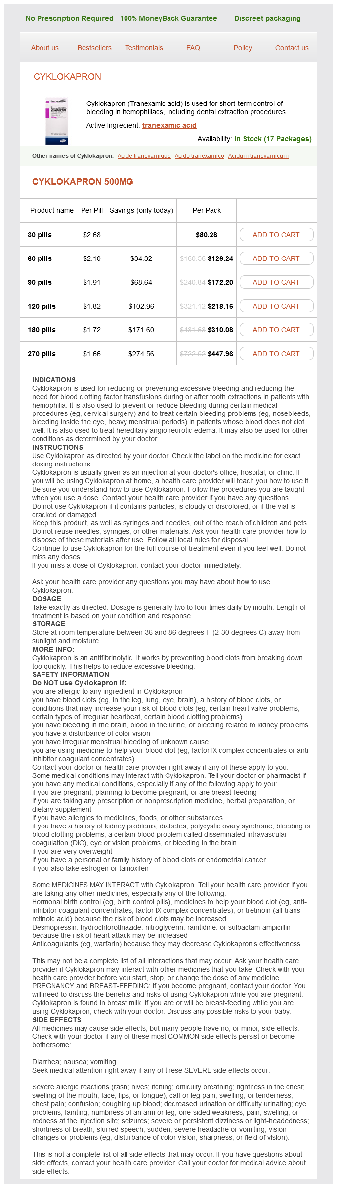
Tranexamic Acid dosages: 500 mg
Tranexamic Acid packs: 30 pills, 60 pills, 90 pills, 120 pills, 180 pills, 270 pills
In stock: 681
Such programs should (1) reverse the underlying physicochemical and physiologic derangements treatment 2014 purchase 500mg tranexamic with mastercard, (2) inhibit new stone formation, (3) overcome nonrenal complications of the disease process, and (4) be free of serious side effects. The rationale for the selection of certain treatments is the One potential criticism of the "selective" metabolic management of nephrolithiasis is that the collection of multiple urine and serum studies can be too time-consuming to be feasible outside of an academic medical center with its dedicated research staff. Although a commitment to follow-up can be tedious, it should be no worse for patients with kidney stones than it is for those followed for urologic cancer or voiding dysfunction. Indeed, Lingeman and colleagues (1998) compared the results of patient management from seven private practices to that achieved by a dedicated university clinic. They found that supersaturation values were effectively reduced in the network and stone clinic and that the reduction was proportional to the initial supersaturation value and increase in urine volume. The stone clinic achieved a greater supersaturation reduction, higher fraction of patient follow-up, and greater increase in urine volume, but the treatment effects in the network were, nevertheless, substantial and significant. This finding is supported by a further study demonstrating the efficacy of medical prophylaxis when administered in a private practice setting (Mardis et al, 2004). When compared to conservative measures of dietary recommendations and fluid management, active pharmacologic treatment achieved a significantly greater reduction in stone episodes. These findings prompted Mardis and coworkers (2004) to conclude that medications validated in trials and guided by metabolic evaluation lower stone recurrence when used in a private practice setting, as they do in clinical trials from academic medical centers. AbsorptiveHypercalciuria Thiazides Currently no treatment program is capable of correcting the basic abnormality of absorptive hypercalciuria I and thiazide diuretics are not considered a selective therapy for absorptive hypercalciuria, because they do not decrease intestinal calcium absorption in this condition (Pak, 1979). However, this class of medication has been widely used to treat absorptive hypercalciuria, because of its hypocalciuric action and the high cost and inconvenience of alternative therapy (sodium cellulose phosphate, which is no longer available in the United States). The use of thiazides was first described by Yendt and colleagues (1966) for the treatment of undifferentiated hypercalciuria. Thiazides directly stimulate calcium resorption in the distal nephron while promoting excretion of sodium. Long-term thia- zide therapy results in volume depletion, extracellular volume contraction, and proximal tubular resorption of sodium and calcium. Thiazides may increase urinary excretion of magnesium and zinc, but these responses are not consistent. Potassium losses from thiazide therapy can cause hypocitraturia, as a result of hypokalemia with intracellular acidosis. Studies indicate that thiazide may have a limited long-term effectiveness in absorptive hypercalciuria type I (Zerwekh and Pak, 1980; Preminger and Pak, 1987). Despite an initial reduction in urinary excretion, the intestinal calcium absorption remains persistently elevated. These studies suggest that the retained calcium may be accreted in bone at least during the first few years of therapy. Bone density, determined in the distal third of the radius by photon absorptiometry, increases significantly during thiazide treatment in absorptive hypercalciuria, with an annual increment of 1. With continued treatment, however, the rise in bone density stabilizes and the hypocalciuric effect of thiazide becomes attenuated. These results suggest that thiazide treatment may cause a low turnover state of bone that interferes with a continued calcium accretion in the skeleton.
Nimbaka (Lemon). Tranexamic Acid.
Source: http://www.rxlist.com/script/main/art.asp?articlekey=96546
The holmium laser can be used to incise the narrow diverticular neck treatment alternatives order tranexamic 500 mg without a prescription, fragment stones within, and ablate the diverticular lining. Stone-free rates of 50% to 90% are found in most series, though Auge and colleagues (2002) found a much lower symptom-free rate of 35% (Fuchs and David, 1989; Grasso et al, 1995b; Batter and Dretler, 1997; Chong et al, 2000; Auge et al, 2002; Legraverend et al, 2013). Adequate diverticular obliteration is lower with the ureteroscopic approach (approximately 20%) than with a percutaneous approach (>70%), hence the need to ensure a patent and well-draining diverticular neck. Unfortunately, the ostium to calyceal diverticulum cannot be successfully located in up to 25% of cases, and when this occurs the diverticular stones cannot be treated ureteroscopi- Horseshoe Kidneys and Renal Ectopia Horseshoe Kidneys. Horseshoe kidneys are the most common renal fusion anomaly, with a reported incidence of 1 in 400 live births (Pitts and Muecke, 1975; Evans and Resnick, 1981). It is important to recognize that there is a 15% to 20% incidence of kidney stone disease in horseshoe kidneys. Most stones are composed of calcium oxalate, with the most common locations being the renal pelvis and posterior lower pole calyces (Evans and Resnick, 1981; Tan et al, 2013). Embryonically, the abnormal medial fusion of the left and right metanephric blastemata creates an isthmus that anchors the fused kidneys at the level of the inferior mesenteric artery, leading to incomplete renal ascent and malrotation (Hohenfellner et al, 1992). These anatomic and functional changes have an impact on the various treatment options for renal stones, and specific horseshoe kidney anatomy, stone location, and stone size must also be considered when choosing the optimal stone treatment. Moreover, multiple treatment sessions are almost always necessary (Lampel et al, 1996; Elliott et al, 2010; Ray et al, 2011; Tan et al, 2013). On average, a higher number of shocks are necessary per treatment session, and a higher re-treatment rate is found versus similar stones in orthotopic, anatomically normal kidneys (Chaussy and Schmiedt, 1984; Drach et al, 1986; Lingeman et al, 1986). Sheir and colleagues (2003) found superior stone-free rates of 79% for stones up to 15 mm, compared with 53% for stones larger than 15 mm. Kirkali and colleagues (1996) similarly found poor stone-free rates (28%) for stones larger than 10 mm. In addition, the more anteriorly and centrally positioned horseshoe kidney causes the access tract to be longer, and this may necessitate use of extralong access sheaths, nephroscopes, and instruments, especially in obese patients. Supracostal access is rarely necessary because the entire horseshoe kidney is often situated below the 12th ribs, and consequently pleural injuries are rare (Raj et al, 2003; Shokeir et al, 2004). Laparoscopic assistance is only rarely used for stone surgery on horseshoe kidneys and only a few case reports exist. Ectopic kidneys are most commonly situated in the pelvis, with the incidence of pelvic kidneys estimated at 1 in 2200 to 1 in 3000 patients. More rarely, ectopic kidneys can be located in the abdomen, in the thoracic cavity, or in a crossed, retroperitoneal location. The approach to kidney stone treatment in these instances should be highly tailored to the specific individual, stone burden, and kidney location, along with any associated kidney drainage impediments. Shock wave lithotripsy achieves stone-free rates of 25% to 92%, although multiple treatment sessions are the norm (Theiss et al, 1993; Talic, 1996; Semerci et al, 1997; Gallucci et al, 2001; Sheir et al, 2003; Tunc et al, 2004). With the pelvic kidney shielded posteriorly by the bony pelvis, prone positioning is often necessary to improve shock wave delivery to the pelvic kidney stones when this technique is selected.
Antibiotics should be given because of the risk for infected urine and abscess Ureteral Access Sheath the use of a ureteral access sheath as an adjunct to ureteroscopy was first reported by Takayasu and Aso in 1974 as a means to simplify access to the intrarenal collecting system medicine 3604 buy tranexamic 500mg on-line. It was not until more than two decades later that the ureteral access sheath was rediscovered and refined, simplifying the deployment and safety of these devices. The present generation of ureteral access sheaths consists of a hydrophilic outer coating, as well as a tapered transition from obturator to sheath, which facilitates their retrograde placement. The walls of the sheaths are designed not only for a slim profile but also for strength and often are reinforced so as to resist kinking. Kourambas and associates (2001) reported that the ureteral access sheath can be successfully deployed in over 90% of attempted placements. In this randomized controlled study these authors also found that the use of an access sheath decreased operating room time, simplified re-entry of the ureter, and, likely as a consequence of these two points, was associated with decreased operating room costs. When the stone migrates only to the submucosa, a problematic complication can develop, because removal of such stones is difficult. If submucosal stones are encountered, laser excision followed by ureteral stent placement is recommended. Submucosal stones are of concern, because they can increase the risk for ureteral stricture formation. Complete extrusion of a calculus, also known as a lost stone, can occur in the setting of a ureteral perforation. In most cases, if the fragment is completely outside the collecting system it can be left in place. Attempts to retrieve the stone may exacerbate the injury and increase the risk for significant irrigant extravasation. When an extruded stone is recognized, the procedure should be terminated and a ureteral stent placed. Antibiotics should be administered to prevent the theoretical risk for abscess formation, although such a complication would be rare. One of the most serious sequelae of such an event is the later development of a ureteral stricture; for this reason patients who have calculus extrusion should undergo postoperative imaging, which will confirm the stone location. It is possible that, in the future, the lost stone could be confused for a ureteral stone, and it is important for the patient to be aware that such a situation exists. Perhaps the most catastrophic complication that can occur during a ureteroscopic procedure is avulsion of the ureter. Ureteral avulsion generally occurs as a consequence of overly forceful manipulation of a large or impacted calculus; however, a scabbard effect also can be created with resulting avulsion at time of scope withdrawal if too large a rigid ureteroscope is forcefully advanced up the ureter.
Syndromes
Additional information:
Usage: b.i.d.
Tags: cheap tranexamic 500 mg on-line, generic tranexamic 500mg line, tranexamic 500 mg, buy 500 mg tranexamic fast delivery
Nefarius, 39 years: Shock wave lithotripsy after pushback into the bladder has been reported, although success rates were only reported at 60% (El-Sharif and Prasad, 1995). Division of the vas deferens should be performed approximately 3 cm distal to the cauda of the epididymis in the straight portion of the vas deferens at the time of vasectomy. In a more recent report, metabolic abnormalities were found in 3 of 5 patients with pure struvite stones and 17 of 22 with mixed struvite stones (Iqbal et al, 2013).
Kaffu, 45 years: More recently, Nguyen and colleagues (2014) described their experience with panurothelial disease. Furthermore, ureteral dilatation has been reported to result from retroperitoneal inflammatory processes secondary to appendicitis, regional enteritis, ulcerative colitis, or peritonitis (Makker et al, 1972). Urinary prothrombin fragment 1 (F1) is a crystal matrix protein named for its resemblance to the F1 degradation product of prothrombin.
Sanford, 60 years: In males, this could include one or both upper urinary tracts and/or the prostatic urethra, and in females the bladder and both upper urinary tracts. The perivesical approach is useful in pediatric patients with a large seminal vesicle cyst so that nephroureterectomy can be performed along with seminal vesiculectomy. A double-J ureteral stent is first placed into the ipsilateral ureter cystoscopically.
Treslott, 65 years: Trigonal function also may be a factor in the prevention of vesicoureteral reflux. With a Gibson, low midline, or Pfannenstiel incision, bladder cuff removal is performed using a transvesical. This effect may be partly because protein intake is higher in affluent people, and stone formation, for some reason, seems to be higher in the economically advantaged.
Volkar, 44 years: Comparison of shockwave lithotripsy outcomes in patients receiving sufentanil or lidocaine spinal anesthesia. However, the findings were not independent of stage and grade in multivariate analysis. For a laparoscopic single-site procedure all trocars are inserted via the perumbilical incision.
Darmok, 47 years: Benign fibroepithelial polyps as a cause of intermittent ureteropelvic junction obstruction in a child: a case report and review of the literature. Acute rejection classically occurs approximately 5 days after an allogeneic organ transplant without immunosuppression. Extracorporeal renal lithotripsy: evolution of residual lithiasis treated with thiazides.
Asaru, 27 years: Home tap water samples from urinary stone patient hospitalizations were compared with controls. Because allantoin is 10 to 100 times more soluble in urine than uric acid, humans are prone to uric acid stone formation. Based on these studies, the use of statins to prevent mortality in dialysis patients is not warranted (Navaneethan et al, 2009a).