-
(786) 502-2173
-
We've gone mobile!
-
Hours: By Appointment Only
Only $2.55 per item
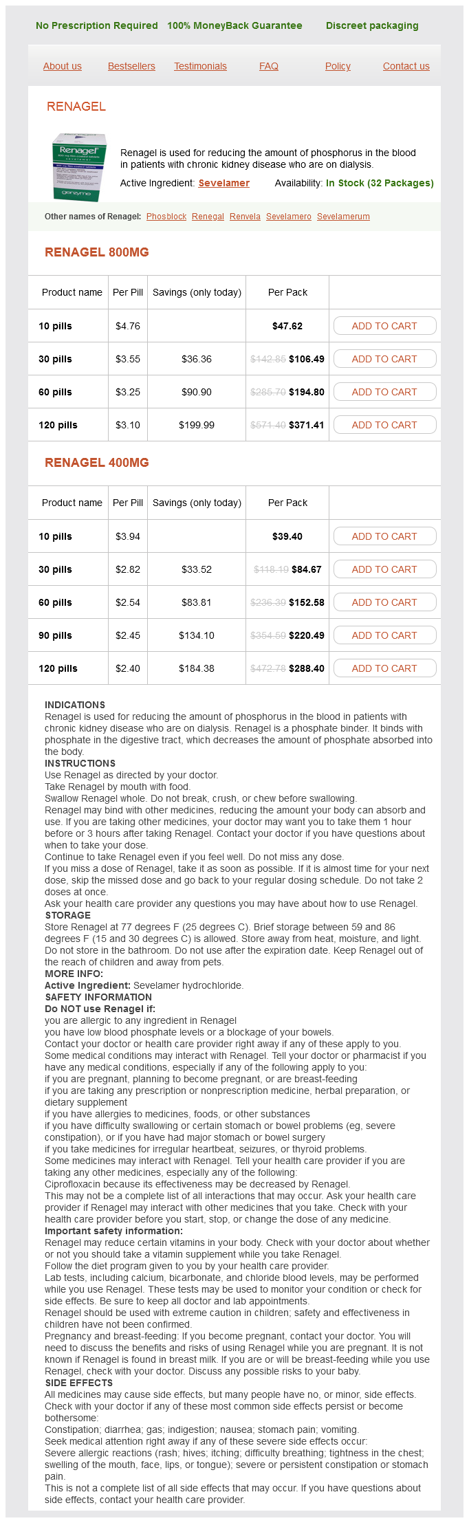
Sevelamer dosages: 800 mg, 400 mg
Sevelamer packs: 10 pills, 30 pills, 60 pills, 120 pills, 90 pills
In stock: 603
These include syncope gastritis diet blog discount sevelamer 400 mg free shipping, breath holding spells, acute psychiatric states, migraine variants, abnormal movement disorders, paroxysmal disturbances of sleep like night terrors, narcolepsy and lastly hysteria. Treatment should be deferred until the diagnosis becomes obvious on followup of the natural course of the disease. Carbamazepine is an effective drug for partial and generalized tonic-clonic seizures. If seizures control is inadequate or toxicity appears, an alternate antiepileptic drug should be tried and the initial drug tapered. Effective agents include ethosuximide, sodium valproate, lamotrigine and benzodiazepines Myoclonic and atonic seizures. Sodium valproate, levetir acetam, benzodiazepines such as clonazepam, nitrazepam or clobazam may be used. Lamotrigine and topiramate have wide spectrum and are useful adjunct after primary failure in partial and generalized epilepsies. Surgical Treatment Medically resistant cases of epilepsy may be treated surgically after a careful selection and work up. Ataxia, nystagmus, diplopia, drowsiness, seizures (phenytoin encephalopathy), rash. Nausea, sedation, weight gain, hair loss Photophobia, leukopenia, drowsiness, nausea. Well oriented (5), confused conversation (4), inappropriate words are spoken (3), incomprehensible sounds (2), no vocal response (1). Diagn osis of Coma functioning of the brainstem and deep coma is unlikely However, flexion and extension movements may be seen in comatose patients as they are mediated at lower spinal reflex level. Bilateralfixed dilated pupils are seen in terminal states or severe ischemic brain damage, atropine or belladonna poisoning. Unilateral unreactive dilated pupils indicate third nerve damage, often associated with transtentorial herniation of the temporal lobe or traction of third cranial nerve against posterior cerebral artery. Stimulation of cortical center for gaze, results in conjugate eye movements to the contralateral side, whereas ablation produces conjugate deviation of the eyes to the ipsilateral side. If the head is suddenly turned to one side, there is a conjugate deviation of eye in the opposite direction indicating that brainstem is intact. If the external auditory canal is irrigated with cold water, the eyes normally deviate towards the stimulated side. This response is lost in pontine lesions, labyrinthitis and coma due to drugs such as sedatives and phenytoin. The hallmark of metabolic encephalopathy consists of loss of oculocephalic and oculovestibular reflexes with preservation of the pupillary light reflex. Structural lesions involving cortical or subcortical motor areas lead to contralateral hemiparesis, hemifacial weakness, partial seizures or tone changes.
Fucostanol (Sitostanol). Sevelamer.
Source: http://www.rxlist.com/script/main/art.asp?articlekey=96834
Between the cerebral hemispheres gastritis and constipation diet order 400 mg sevelamer, the anterior cerebral arteries are cut in cross section as they ascend from the circle of Willis in the region of the sella turcica to the anterior cerebrum. Near the center of the image, the third ventricle appears as a clearly distinct radiolucent area between the thalamic nuclei, which are surrounded by the internal capsules. Although spots of high density appear to be within the third ventricle, they are calcifications within the pineal gland found outside the third ventricle between the quadrigeminal plate and the splenium of the corpus callosum. Although the radiolucent area between the pineal body and the cerebellar vermis appears to be the same density as the ventricle previously described, this area is formed by an enlarged part of the subarachnoid space outside of the brain, the superior cistern. Forming the border between the occipital lobes of the cerebrum and the upper part of the cerebellum, the tentorium cerebelli is sectioned, demonstrating the straight sinus and the confluence of sinuses that are formed in part by an extension of dura mater from the tentorium cerebelli. Anteriorly, the heads of the caudate nuclei are shown protruding in to the anterior horns of the lateral ventricles. Between the cerebral hemispheres, the contrastenhanced anterior cerebral arteries are shown in cross section as they extend from the base of the brain where they originate from the circle of Willis to extend upward to supply blood to the anterior cerebral hemispheres. At this level, the midline ventricle previously identified as the third ventricle is sectioned near the top of the opening. As described previously, the lateral walls of the third ventricle are formed by the thalamic nuclei. On the opposite side of the thalamic nuclei, the internal capsules act to separate the thalamic nuclei from the basal ganglia: caudate nucleus, globus pallidus, and putamen. Posterior to the thalamic nuclei, the tails of the caudate nuclei are shown in cross section as they extend toward the inferior horns of the lateral ventricles. In the midline between the cerebral hemispheres, the vein of Galen is shown in cross section as a large contrast-enhanced vessel directly behind the third ventricle. The vein of Galen drains venous blood in to the straight sinus obliquely sectioned between the posterior cerebral hemispheres. Similar to previous images, the anterior horns of the lateral ventricles are readily identified as radiolucent areas within the anterior cerebral hemispheres. The anterior horns of the lateral ventricles are separated by the septum pellucidum, which extends between the splenium and genu of the corpus callosum. Between the anterior and posterior horns of the lateral ventricles, the contrast-enhanced choroid plexus lies within the bodies of the lateral ventricles. Similar to the previous image, the contrast-enhanced vein of Galen and straight sinus are sectioned between the posterior cerebral hemispheres. On either side, the ventricles are surrounded by an area of white matter formed primarily of neural fibers extending to and from the gray matter of the cerebral cortex. Just medial to the lateral ventricles, the region of white matter represents the body of the corpus callosum and consists of a group of nerve fibers extending between the right and left cerebral hemispheres. In the midline, the falx cerebri is formed by a reflection of dura mater separating the right and left cerebral hemispheres. Similar to the previous image, the falx cerebri is shown separating the right and left cerebral hemispheres. Along its margin, the falx cerebri forms the superior sagittal sinus and is labeled on the posterior part of the image adjacent to the parietal bone.
Similar to previous images gastritis que no comer sevelamer 800 mg purchase otc, the ureters appear as bright, contrast-enhanced vessels near the psoas muscles and are in close proximity to the major vessels of the pelvis. In the anterior pelvic cavity, numerous loops of small bowel are loosely organized centrally, and the descending colon is seen on the left side occupying a position near the iliacus muscle. Lining the posterior pelvic wall, the iliacus and psoas muscles are shown in cross section adjacent to the iliac bones. In this image, the external and internal iliac arteries are shown on either side of the left ureter and in front of the larger common iliac vein. Like previous images, the descending colon and the cecum are on opposite sides of the randomly organized loops of small bowel distributed within the peritoneal cavity. Although the first pair of sacral foramina is near the point of exit from the sacrum, the second pair is just emerging from the sacral canal. On either side of the sacrum, the iliac bones form the sacral iliac joints where they articulate with the lateral parts of the sacrum. Within the greater pelvis, the left ureter is enhanced and lies near the medial border of the psoas muscle. Within the peritoneal cavity, the loops of small bowel occupy most of the anterior pelvis. Because this part of the small bowel is within the pelvis, the ileum is probably the part of the small bowel shown. Common iliac A 416 Introduction to Sectional Anatomy the sacrum, iliac bones, iliacus muscles, and psoas muscles are shown in cross section forming the posterior wall of the pelvic cavity. Compared to previous images, the psoas and iliacus muscles are not as clearly separable as they join to form the iliopsoas muscles in the lower pelvis. Near the psoas muscles, the ureters are readily visible as contrast-enhanced structures between the external and internal iliac vessels. The intestinal structures are similar to previous views, with the small bowel occupying most of the peritoneal cavity. Posterior to the peritoneum, the lower edge of the cecum is shown on the right and the descending colon is demonstrated on the left. Although the descending colon ends in the sigmoid colon, the irregular shape of the sigmoid colon moves upward so that it is also included within this section. Lt common iliac A 418 Introduction to Sectional Anatomy the sacrum is smaller than in the previous images. Although the ilium is shown in cross section similar to previous views, the central portion of the ilium is beginning to expand, indicating that the section is nearing the acetabulum. Within the pelvic cavity, the sigmoid colon is more clearly seen in its characteristic S shape as it extends toward the descending colon on the left side.
Syndromes
Additional information:
Usage: q.d.
Tags: 800 mg sevelamer, buy sevelamer 800 mg with mastercard, order sevelamer 800 mg with amex, buy 800 mg sevelamer with visa
Grok, 31 years: Several studies have reduced the dose of radiation in patients achieving a favorable response to chemotherapy. Migraine and other primary headache disorders may only be diagnosed in retrospect after their episodic course becomes manifest and there is no evidence of an underlying lesion.
Kafa, 24 years: Eight-hundred milliliters of normal saline is infused in to the right pleural cavity. Epidemiologic studies provide information about the rates and risk factors for the onset, progression, and remission of migraine.
Narkam, 52 years: Treatment of intrahepatic cancers with radiation doses based on a normal tissue complication probability model. As noted before, headaches secondary to organic disease or dysfunction must be ruled out before a diagnosis of primary headache disorder is established.
Yorik, 46 years: Blunt crushing of the hepatic parenchyma exposes biliary and vascular structures, which may then be divided between ligature clips. It is often helpful to begin with the current headache of greatest concern to the patient and subsequently explore other headache patterns and the evolution of those patterns.
Hanson, 39 years: These drugs should be discontinued at the first sign of tachyarrhythmia, hypotension or diminished systemic vascular resistance. It is successful in cases of sudden onset fibril lation along with oxygenated normothermic myocardium without significant acidosis.