-
(786) 502-2173
-
We've gone mobile!
-
Hours: By Appointment Only
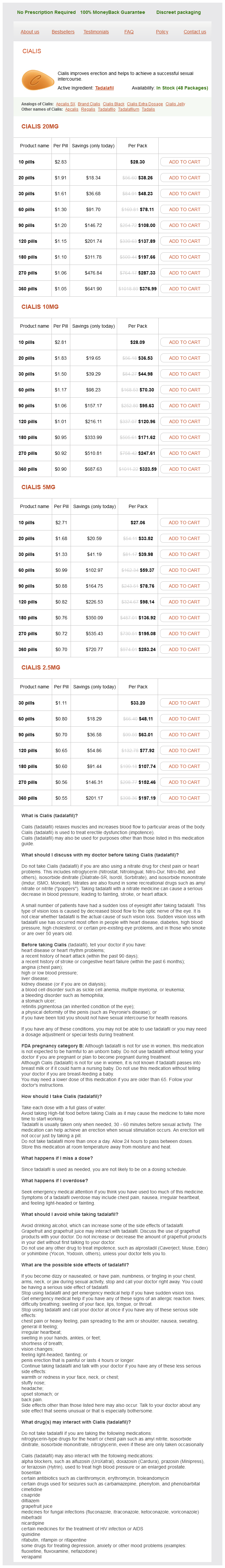
Only $0.58 per item
Regalis dosages: 20 mg, 10 mg, 5 mg, 2.5 mg
Regalis packs: 10 pills, 20 pills, 30 pills, 60 pills, 90 pills, 120 pills, 180 pills, 270 pills, 360 pills
In stock: 658
Cardiac axis angles outside this range indicate possible cardiac abnormalities erectile dysfunction drugs and heart disease regalis 2.5 mg lowest price, possibly due to a mass effect from intrathoracic masses or diaphragmatic hernia, or other abnormalities. Crane found cardiac axis outside of the normal range as 79% sensitive and 97% specific for congenital cardiac abnormalities, and 5 of 23 infants with structural cardiac anomalies and an abnormal axis were initially thought to have a normal four-chamber view of the heart (Crane et al. Cardiac axis to the right is rare but strongly associated with underlying cardiac abnormalities. Comstock found 22 such infants among 16,562 fetuses evaluated over a 6-year period. Twelve infants had isolated rotation of the heart axis, six fetuses had mirror-image hearts with situs inversus, and four had inversion of the ventricles. Fourteen of the 22 infants had structural cardiac defects, most of which were atrioventricular septal defects, double outlet right ventricles, or common atria. Neonatal outcomes were good in 16 of the infants, while 4 infants with polysplenia or asplenia, and 2 infants with other severe extracardiac malformations died (Comstock et al. The membranous septal portion of the intraventricular septum, located immediately adjacent to the cardiac crux, is anatomically very thin. If imaged in anything other than an ideal imaging plane, the membranous septum may appear discontinuous, having a tendency to simulate a membranous ventricular septal defect. Further imaging from slightly different angles and evaluation with color Doppler color flow or pulsed Doppler will usually exclude such false positive diagnoses. Small ventriculoseptal defects may also go undetected, but this is often of minimal clinical significance, as small defects often resolve spontaneously during the first months and years of life. These include the left ventricular outflow view and short axis great vessel views, which are usually obtained by rotating the longitudinal axis of the transducer from the fourchamber view by 30 45 to the right or left, resulting in imaging planes that transect the scapula and torso at 30 45 angles from the midsagittal plane. In addition, the five-chamber view is a modification of the four-chamber imaging plane in which the aortic root origin in the left ventricle is imaged (one chamber), as well as accompanying views of the other four standard chambers giving a five-chamber appearance. The left ventricular outflow tract view shows left atrium, left ventricle, the mitral valve, and the aorta. The right ventricular outflow tract view shows the right ventricle, the pulmonic valve, the pulmonary artery, and a portion of the ductus arteriosus. A crossing view of the pulmonary artery and aorta in which the pulmonary artery and right outflow tract can often be shown to cross the aorta and left ventricular outflow tract in a transverse fashion at an angle of about 30 generally excludes the transposition of the great vessels. The short axis great vessel view demonstrates the right cardiac structures (right atrium, tricuspid view, right ventricle, and pulmonic valve) arrayed circumferentially around the aortic root, with the bifurcation of the pulmonary artery into the ductus and the right pulmonary artery clearly seen. The triple leaf pattern of the aortic valve (resembling the letter Y) is often seen. This view is helpful to show the discrete origin of the major great vessels and assists with evaluation of complex cardiac abnormalities.
Trigonella Foenum-Graecum (Fenugreek). Regalis.
Source: http://www.rxlist.com/script/main/art.asp?articlekey=96717
Increased placental resistance results in high umbilical S/D ratios and increased pulsatility erectile dysfunction rap beat regalis 20 mg without a prescription. Conversely, redistribution of cerebral flow in fetal growth restriction is often associated with lower cerebral artery S/D ratios and less pulsatility then is found in normal infants. Evaluations of flow in the cerebral circulation require relatively high-quality Doppler equipment, but such equipment is becoming increasingly more readily available as sonographic equipment becomes more advanced and prices decrease. The reconstructed images can then be rotated and viewed from many different aspects. The ability to manipulate the images in this fashion allows certain structural anomalies such as neural tube anomalies and facial clefts to be viewed much more easily and from more favorable angles. They also offer the advantage of generating an image that is much more easily understood and interpreted by the lay public and that facilitates understanding of the high degree of anatomic disarrangement often present in severe anomalies. Routine Antepartum Sonography It is estimated that basic ultrasonography is performed in 6070% of pregnancies, but in the United States, sonography of low-risk patients has never been established as a requisite of routine care (Dooley 1999). This may relate to the relatively low incidence of anomalies, the rather poor sensitivity of ultrasound screening for identifying structural anomalies as described below, and the limited options available for correction of the problems identified. Additionally, many clinical situations, such as multiple gestations, excessive fetal growth, fetal growth restriction, oligohydramnios, and polyhydramnios will present clinical findings that initiate a process of evaluation resulting in their diagnosis by ultrasound evaluation. Although antepartum sonographic screening has been found associated with lower perinatal mortality at term, most of that reduction has been ascribed to voluntary terminations of infants with severe anomalies that otherwise would be included in perinatal mortality statistics. In England, patients often undergo an initial sonographic evaluation at the time of their first prenatal visit to confirm menstrual dating, followed by an anatomic survey in the early second trimester, and a sonogram to confirm normal fetal growth and development in the early third trimester. Although, such utilization patterns may not yield statistically improved morbidity and mortality statistics, they afford unquestionable opportunities for parents to bond with their children, identify fetal growth restriction before it is clinically evident, and in cases of major anomalies, may offer the advantages of more prompt prenatal diagnosis. Indications for Antenatal Sonography As with other tests, sonography is usually performed to answer a clinical question such as excluding the possibility of ectopic gestation, surveying for structural anomalies or abnormalities of placentation, assessing size/gestational age disparities (as might occur if the gestational age has been incorrectly assessed, in cases of multiple gestation, with aberrant fetal growth, or if the amniotic fluid volume is abnormal), or to evaluate fetal well-being. Although fetal size can be estimated clinically to a reasonable degree of accuracy in low-risk pregnancies, this is sometimes impossible (in multiple gestation) or impractical (in cases of morbid maternal obesity). Serial sonographic estimates in these circumstances are more sensitive early markers of aberrant fetal growth than clinical size assessment. Also, newer Doppler sonographic techniques can be used to document circulatory changes associated with ongoing fetal nutritional stress such as the shift of fetal circulation to the cerebral vasculature in growth restriction and the increased flow velocity in the middle cerebral artery associated with fetal anemias due to rhesus and other atypical forms of isoimmunization. Indications for sonography include the following: Estimation of gestation age in patients with uncertain clinical dates, who plan to deliver by indicated induction of labor or scheduled repeat elective cesarean, or who plan to electively terminate their pregnancies. Initially, well-being assessment centered on sonographic evaluations of amniotic fluid volume after increased rates of morbidity and mortality were noted in post-term fetuses with abnormally low amniotic fluid volume. Criteria such as absence of a fluid pocket greater than 1 cm2 or 2 cm2 eventually gave way to assessments of the amniotic fluid index, computed as the sum of the deepest amniotic fluid pocket from the four quadrants of the amniotic cavity. Amniotic fluid indices were then evaluated as falling above or below certain critical values (values < 5 or 6 often characterized as oligohydramnios, values >25 considered polyhydramnios) or assessed with gestationally specific norms (Moore and Cayle 1990). Sonography also allows the possibility of dynamic, real-time assessment of the fetus. The sonographic criteria to be evaluated over a 30-min interval included presence of fetal breathing movements of 30 seconds or greater, fetal tone, gross body movements, and presence of a normal amniotic fluid volume (various criteria have been used over time).
Acute and chronic desensitization of penicillin-allergic patients using oral penicillin erectile dysfunction 47 years old regalis 10 mg line. Vancomycin hypersensitivity: Synergism with narcotics and "desensitization" by a rapid continuous intravenous protocol. Vancomycin anaphylaxis and successful desensitization in a patient with end stage renal disease on hemodialysis by maintaining steady antibiotic levels. Rapid imipenem/cilastatin desensitization for multidrug-resistant Acinetobacter pneumonia. Antibiotic desensitization for the allergic patient: 5 years of experience and practice. Effective acute desensitization for immediate-type hypersensitivity to human granulocyte-monocyte colony stimulating factor. Case reports of evaluation and desensitization for anti-thymocyte globulin hypersensitivity. Desensitization of patients allergic to penicillin using orally administered beta-lactam antibiotics. Rapid inpatient/outpatient desensitization for chemotherapy hypersensitivity: standard protocol effective in 57 patients for 255 courses. Efficacy and safety of desensitization to allopurinol following cutaneous reactions. Aspirin desensitization in aspirin-sensitive asthmatic patients: clinical manifestations and characterization of the refractory period. Long-term treatment with aspirin desensitization in asthmatic patients with aspirinexacerbated respiratory disease. Aspirin desensitization treatment of aspirin-sensitive patients with rhinosinusitisasthma: long-term outcomes. The blocking effect of essential controller medications during aspirin challenges in patients with aspirin-exacerbated respiratory disease. Aspirin desensitization in patients undergoing percutaneous coronary interventions with stent implantation. Rapid oral challengedesensitization for patients with aspirin-related urticaria-angioedema. Rapid desensitization procedure for patients with aspirin hypersensitivity undergoing coronary stenting. Routine elective penicillin allergy skin testing in children and adolescents: study of sensitization. Natural evolution of skin test sensitivity in patients allergic to beta-lactam antibiotics.
Syndromes
Additional information:
Usage: ut dict.
Tags: generic 20 mg regalis mastercard, purchase 5 mg regalis with amex, order regalis 20 mg mastercard, order 20 mg regalis otc
Jack, 47 years: Physiological parameters of morphine dependence in man tolerance, early abstinence and protracted abstinence. Rarely, a central web within the stomach may obstruct flow out of the stomach, leaving a single bubble 4. The cerebellar tonsils will be approximated with mild to moderate edema and will be herniated through the foramem magnum when there is severe edema.
Givess, 42 years: Detection of IgE antibodies to bacitracin using a commercially available streptavidin-linked solid phase in a patient with anaphylaxis to triple antibiotic ointment. A comparison of the efficacy and safety of olanzapine versus haloperidol during transition from intramuscular to oral therapy. Biopsy-proven eosinophilic pneumonia may occur after use of sulfonamides, penicillin, and para-aminosalicylic acid.
Rozhov, 43 years: Biologic agents for the treatment of juvenile rheumatoid arthritis: current status. Amniotic fluid before 1618 weeks gestation is usually present as a result of transfer of fluid across the placental membranes and does not give helpful information about renal function. A 4-week, multicenter, double-blind, placebo-controlled trial of pregabalin and alprazolam.