-
(786) 502-2173
-
We've gone mobile!
-
Hours: By Appointment Only
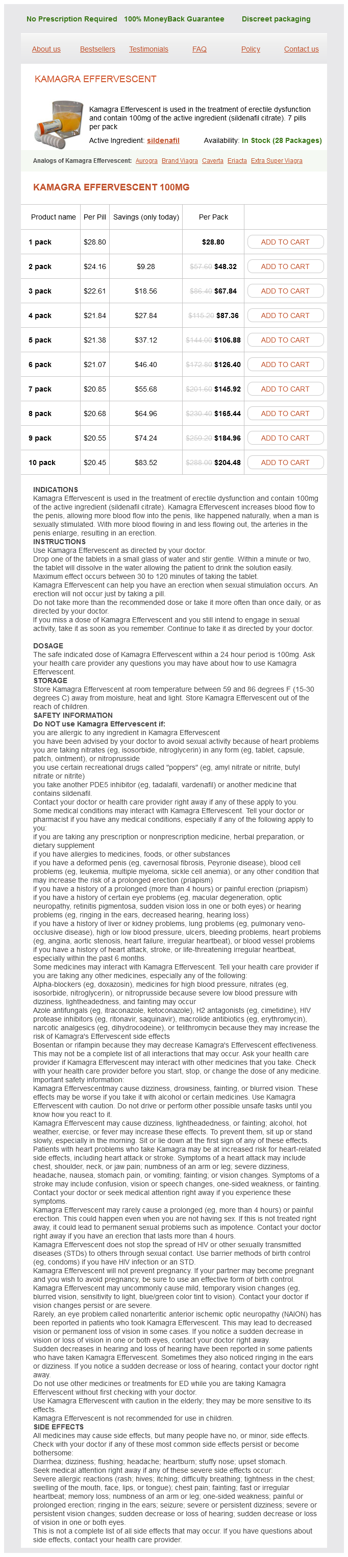
Only $21.73 per item
Kamagra Effervescent dosages: 100 mg
Kamagra Effervescent packs: 1 pack, 2 pack, 3 pack, 4 pack, 5 pack, 6 pack, 7 pack, 8 pack, 9 pack, 10 pack
In stock: 712
Imaging demonstrates a smooth segment of ureteral narrowing with or without associated obstruction erectile dysfunction pump.com generic 100 mg kamagra effervescent free shipping. Crosssectional imaging is the most helpful imaging modality because it shows bowel inflammation and the extent of the inflammatory process with a soft tissue mass or fistula involving the ureter. In the setting of diverticulitis, which commonly involves the sigmoid colon, the left ureter is usually involved. The ureter may be displaced or compressed by an adjacent abscess or encased by inflammation. Rarely, complications from appendicitis, such as abscess formation and inflammation, can extend to involve the right ureter. The smooth segmental ureteral narrowing seen with inflammatory bowel processes that involve the ureter are similar to that in other benign ureteral strictures. Depending on the cause and severity of ureteral involvement, management may involve stent placement or surgical intervention. Endometriosis Involvement of the ureter by endometriosis is usually seen in the setting of extensive pelvic disease and occurs in 0. Patients with endometriosis usually present with pelvic pain associated with menses and infertility. When the ureters are involved, patients may present with hematuria and flank pain that may occur at the time of menstruation as well as obstruction. In endometriosis, ectopic endometrial tissue implants on the peritoneal surfaces of the pelvis. Hormonal changes of the menstrual cycle result in repeated episodes of hemorrhage from the endometriotic implants with resultant inflammation and fibrosis. Common sites include the ovaries and fallopian tubes, uterine ligaments, peritoneal reflections, sigmoid colon, and bladder. Endometriosis may cause ureteral narrowing by extrinsic compression by transmural invasion from adjacent endometrial implants or can incite surrounding fibrosis and smooth muscle proliferation. Imaging findings depend on the size of the endometrial implants and the extent of ureteral involvement. Although it often shows adnexal involvement, ureteral implants are not well demonstrated. However, the signal intensity of implants is variable owing to the different age and stages of blood products and the different types of implants. Some implants consist mostly of glandular tissue, are T2 hyperintense, and enhance after administration of a contrast agent. Solid endometriosis has hyperintense foci of hemorrhage as well as surrounding fibrosis, which is hypointense on T2-weighted images and enhances. In the setting of deep pelvic endometriosis, endometrial implants and fibrosis can involve the ureter, resulting in ureteral stricture and obstruction. The imaging appearance of ureteral endometriosis cannot be differentiated from malignant urothelial lesions. Identification of endometriosis in the absence of other pelvic disease or manifestations associated with other entities assists in making the diagnosis.
Chirca Melosa (Carqueja). Kamagra Effervescent.
Source: http://www.rxlist.com/script/main/art.asp?articlekey=97071
The appendages erectile dysfunction caused by vyvanse generic 100 mg kamagra effervescent with mastercard, which often have narrow pedicles, consist of skin, but they may contain some cartilage. Absence of Auricle Anotia (absence of the auricle) is rare but is commonly associated with the first pharyngeal arch syndrome. This defect often serves as an indicator of associated birth defects, such as atresia of the external acoustic meatus (80% of cases) and middle ear anomalies. The sinuses are usually narrow tubes or shallow pits that have pinpoint external openings. Preauricular sinuses may be associated with internal anomalies such as deafness and kidney malformations. The embryologic basis of auricular sinuses is uncertain, but it may relate to incomplete fusion of the auricular hillocks or to abnormal mesenchymal proliferation and defective closure of the dorsal part of the first pharyngeal groove. Most of this pharyngeal groove normally disappears as the external acoustic meatus forms. Other auricular sinuses appear to represent ectodermal folds that are sequestered during formation of the auricle. The preauricular sinus usually is unilateral and involves the right side, and bilateral preauricular sinuses are typically familial. Most sinuses are asymptomatic and have only minor cosmetic importance; however, they can become infected. The deep part of the meatus is usually open, but the superficial part is blocked by bone or fibrous tissue. The auricle is also severely affected, and middle and internal ear defects sometimes occur. Atresia of the external acoustic meatus can occur bilaterally or unilaterally and usually results from inheritance of an autosomal dominant trait. Congenital Cholesteatoma A congenital cholesteatoma is a fragment of keratinized epithelial cells that is retained after birth. The embryonic rest remnants form epithelial tissue that appears as a white, cyst-like structure medial to and behind the tympanic membrane. There is no opening of the external acoustic meatus, but computed tomography revealed normal middle and internal ear structures. Notice the external orifice of the fistula below the auricle, the upward direction of the catheter (in the sinus tract) toward the external acoustic meatus, and the normal position of the auricle. The grooves form at the beginning of the fourth week and deepen to form hollow optic vesicles that project from the forebrain. The optic vesicles contact the surface ectoderm and induce development of the lens placodes. As the lens placode thickens to form a lens pit and lens vesicle, the optic vesicle invaginates to form the optic cup. The sphincter and dilator muscles of the iris develop from the ectoderm at the rim of the optic cup. The surface ectoderm gives rise to the lens and the epithelium of the lacrimal glands, eyelids, conjunctiva, and cornea.
Congenital hernias include indirect inguinal hernias and gastroschisis and omphalocele fast facts erectile dysfunction buy kamagra effervescent 100 mg with mastercard, occurring lateral to or at the umbilicus, respectively. Imaging In the past, the diagnosis of a hernia was made clinically or by means of plain radiographs or barium studies. In some cases, such as in patients with a large wall defect or large hernia sac, the hernia may be visible. In some cases of complicated abdominal wall hernia, such as bowel incarceration or strangulation, conventional radiographs allow detection of signs of mechanical ileus, bowel loop enlargement, thickening of intestinal folds and air/fluid levels. Abdominal wall hernias represent a frequent imaging finding in the abdomen; thus, their actual prevalence and distribution are probably underestimated in the published literature. They usually manifest at points of weakness of the abdominal wall, where no muscle is present, along the midline, in the linea semilunaris on each side, and in the inferior lumbar space. Note the C-shaped, thick-walled, herniated small bowel loops associated with moderate mesenteric fat stranding. Also note the contralateral displacement of the inferior epigastric vessels (arrowhead). Some authors advocate image acquisition while the patient performs the Valsalva maneuver to increase intra-abdominal pressure, which may aid in demonstrating some hernias, particularly those involving the ventral abdominal wall. Moreover, three-dimensional information and multiplanar reformatted images provide excellent anatomic depiction of abdominal wall anatomy and useful information for surgical planning. Note the air-filled colonic loops herniating into the subcutaneous tissue (arrowheads) through a paraumbilical defect. Limitations Not useful for small defects Ionizing radiation Expensive and not widely available Limited spatial resolution Limited on postoperative patients Patient-limiting factors. However, when necessary, it requires flotation pads to achieve best resolution and avoid the "bang effect" of direct transducer placement on the skin. However, its utility in obese patients or in patients with abdominal wall scarring may be limited. The hernia sac protrudes through the internal inguinal ring, located lateral to the epigastric vessels. They may occur in children (indirect most common) and adults (direct and indirect) and manifest medial (direct) or lateral (indirect) to the inferior epigastric vessels. In children, most inguinal hernias develop because the peritoneal extension accompanying the testis fails to obliterate. In adults, they are caused by acquired weakness and dilation of the internal inguinal ring,4 which is a defect in the fascia transversalis. In females, an indirect inguinal hernia follows the round ligament into the labia majora. In some cases, indirect herniated content may pass all the way into the scrotum (known as a complete hernia) and may contain intestine (small bowel or colon), mesenteric fat, the appendix, foreign bodies, bladder, ureter, or any peritoneal cavity content (fluid, air). Direct inguinal hernias are located medial to the inferior epigastric vessels, are thought to be acquired, appear between 30 and 40 years of age, and are often bilateral.
Syndromes
Additional information:
Usage: gtt.
Tags: buy 100 mg kamagra effervescent fast delivery, order kamagra effervescent 100 mg without prescription, kamagra effervescent 100 mg order visa, order 100 mg kamagra effervescent mastercard
Delazar, 63 years: The advent of genetically modified mice that express fluorescent proteins revolutionized cell lineage and mapping studies allowing high-resolution live visualization of morphogenetic events both in situ and in cultured organ explants. Observe the development of the kidney: ureter, renal pelvis, calices, and collecting tubules.
Marlo, 48 years: Patients taking angiotensin-converting enzyme inhibitors or angiotensin receptor blockers usually show a degree of suppression of aldosterone secretion, reflected in a modest (0. In addition, the downregulation of Y1 receptors, by reducing vascular constriction, could contribute to reductions in vascular resistance in the coronary and renal circulations.
Kalan, 33 years: With a patent foramen ovale and patency of the ductus arteriosus, there is some mixing of blood. The tunica albuginea appears as low signal intensity on T1- and T2-weighted images.
Steve, 60 years: Foremost of these is a failure to improve the serum [Na+], despite reasonable attempts at fluid restriction, or the presence of clinical characteristics associated with poor responses to fluid restriction (see Table 16. The main body of the molecule, the substrate-binding cleft, and the prosegment are shown.
Kaffu, 52 years: Once the acute stage of hypervolemia has been controlled, therapy must be directed toward the prevention or minimization of further acute episodes and improvement in overall prognosis. The right umbilical vein disappears during the seventh week, leaving the left umbilical vein as the only vessel carrying well-oxygenated blood from the placenta to the embryo.