-
(786) 502-2173
-
We've gone mobile!
-
Hours: By Appointment Only
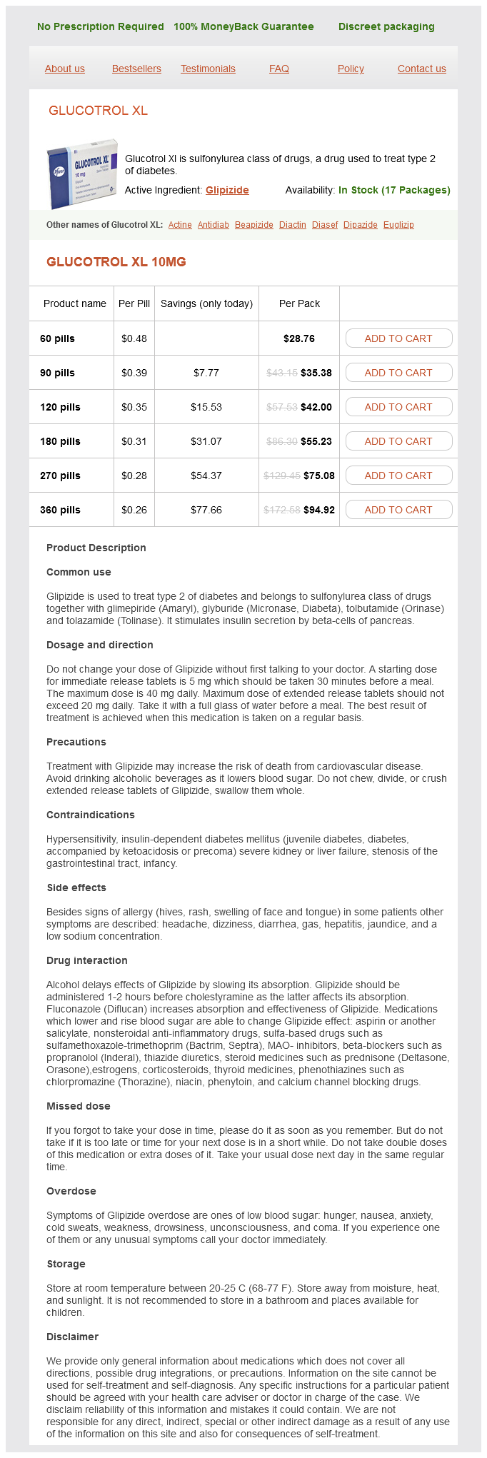
Only $0.28 per item
Glipizide dosages: 10 mg
Glipizide packs: 60 pills, 90 pills, 120 pills, 180 pills, 270 pills, 360 pills
In stock: 646
The left seminal gland and ampulla of the ductus deferens are dissected free and sliced open diabetes type 1 job restrictions order glipizide 10 mg free shipping. The perineal membrane lies between the external genitalia and the deep part of the perineum (anterior recess of ischio-anal fossa). It is pierced by the urethra, ducts of the bulbo-urethral glands, dorsal and deep arteries of the penis, cavernous nerves, and the dorsal nerve of the penis. The superior ends of the seminal glands are covered with peritoneum and lie posterior to the ureters, where the peritoneum of the rectovesical pouch separates them from the rectum. The duct of the seminal gland joins the ductus deferens to form the ejaculatory duct. Although the ejaculatory ducts traverse the glandular prostate, prostatic secretions do not join the seminal fluid until the ejaculatory ducts have terminated in the prostatic urethra. The glandular part makes up approximately two thirds of the prostate; the other third is fibromuscular. The fibrous capsule of the prostate is dense and neurovascular, incorporating the prostatic plexuses of veins and nerves. All of this is surrounded by the visceral layer of the pelvic fascia, forming a fibrous prostatic sheath that is thin anteriorly, continuous anterolaterally with the puboprostatic ligaments, and dense posteriorly where it blends with the rectovesical septum. The anterior surface is separated from the pubic symphysis by retroperitoneal fat in the retropubic space. Lobules and zones of prostate demonstrated by anatomical section and ultrasonographic imaging. The ducts of the glands in the peripheral zone open into the prostatic sinuses, whereas the ducts of the glands in the central (internal) zone open into the 1404 prostatic sinuses and the seminal colliculus. The isthmus of the prostate (commissure of prostate; historically, the anterior "lobe") lies anterior to the urethra. It is fibromuscular, the muscle fibers representing a superior continuation of the external urethral sphincter muscle to the neck of the bladder, and contains little, if any, glandular tissue. Right and left lobes of the prostate, separated anteriorly by the isthmus and posteriorly by a central, shallow longitudinal furrow, may each be subdivided for descriptive purposes into four indistinct lobules defined by their relationship to the urethra and ejaculatory ducts and-although less apparent -by the arrangement of the ducts and connective tissue: 1. An inferoposterior (lower posterior) lobule that lies posterior to the urethra and inferior to the ejaculatory ducts. This lobule constitutes the aspect of the prostate palpable by digital rectal examination. An inferolateral (lower lateral) lobule directly lateral to the urethra, forming the major part of the right or left lobe.
Scilla indica (Squill). Glipizide.
Source: http://www.rxlist.com/script/main/art.asp?articlekey=96725
Although it lies near the center of the diaphragm metabolic brain disease journal impact factor generic 10 mg glipizide with visa, the central tendon is closer to the anterior part of the thorax. The surrounding muscular part of the diaphragm forms a continuous sheet; however, for descriptive purposes, it is divided into three parts, based on the peripheral attachments: Sternal part: consisting of two muscular slips that attach to the posterior aspect of the xiphoid process; this part is not always present. Costal part: consisting of wide muscular slips that attach to the internal surfaces of the inferior six costal cartilages and their adjoining ribs on each side; the costal parts form the right and left domes. Lumbar part: arising from two aponeurotic arches, the medial and lateral arcuate ligaments, and the three superior lumbar vertebrae; the lumbar part forms right and left muscular crura that ascend to the central tendon. The right crus, larger and longer than the left crus, arises from the first three or four lumbar vertebrae. Because it lies to the left of the midline, it is surprising to find that the esophageal hiatus is a formation in the right crus; however, if the muscular fibers bounding each side of the hiatus are traced inferiorly, it will be seen that they pass to the right of the aortic hiatus. The right and left crura and the fibrous median arcuate ligament, which unites them as it arches over the anterior aspect of the aorta, form the aortic hiatus. The diaphragm is also attached on each side to the medial and lateral arcuate ligaments. The medial arcuate ligament is a thickening of the fascia covering the psoas major, spanning between the lumbar vertebral bodies and the tip of the transverse process of L1. The lateral arcuate ligament covers the quadratus lumborum muscles, continuing from the L12 transverse process to the tip of the 12th rib. The superior aspect of the central tendon of the diaphragm is fused with the inferior surface of the fibrous pericardium, the strong, external part of the 1254 fibroserous pericardial sac that encloses the heart. Vessels and Nerves of Diaphragm the arteries of the diaphragm form a branch-like pattern on both its superior (thoracic) and inferior (abdominal) surfaces. The arteries supplying the inferior surface of the diaphragm are the inferior phrenic arteries, which typically are the first branches of the abdominal aorta; however, they may arise from the celiac trunk. Some veins from the posterior curvature of the diaphragm drain into the azygos and hemi-azygos veins (see Chapter 4, Thorax). The veins draining the inferior surface of the diaphragm are the inferior phrenic veins. The anterior and posterior diaphragmatic lymph nodes are on the superior surface of the diaphragm. Lymph from these nodes drains into the parasternal, posterior mediastinal, and phrenic lymph nodes. Lymphatic vessels from the inferior surface of the diaphragm drain into the anterior diaphragmatic, phrenic, and superior lumbar (caval/aortic) lymph nodes.
It is palpable as a soft mass inferior to the body of the mandible blood sugar goals glipizide 10 mg buy, especially when the apex of the tongue is forced against the maxillary incisor teeth. The most common type of torticollis (wry neck) results from a fibrous tissue tumor (L. Spasmodic Torticollis 2267 Cervical dystonia (abnormal tonicity of the cervical muscles), commonly known as spasmodic torticollis, usually begins in adulthood. Characteristics of this disorder are sustained turning, tilting, flexing, or extending of the neck. The shoulder is usually elevated and displaced anteriorly on the side to which the chin turns. Central lines are inserted to administer parenteral (venous nutritional) fluids and medications and to measure central venous pressure. The needle punctures the skin inferior to the thumb (middle of the clavicle) and is advanced medially toward the tip of the index finger (jugular notch) until the tip enters the right venous angle, posterior to the sternoclavicular joint. Here, the internal jugular and subclavian veins merge to form the brachiocephalic vein. If the needle is not inserted carefully, it may puncture the pleura and lung, resulting in pneumothorax. Furthermore, if the needle is inserted too far posteriorly, it may enter the subclavian artery. When the needle has been inserted correctly, a soft, flexible catheter is inserted into the subclavian vein, using the needle as a guide. This vein is not ideal for catheterization because its angle of junction with the subclavian vein makes passage of the catheter difficult. This action produces a churning noise in the thorax and cyanosis (a bluish discoloration of the skin and mucous membranes resulting from an excessive concentration of reduced hemoglobin in the blood). A venous air embolism produced in this way will fill the right side of the heart with froth, which nearly stops blood flow through it, resulting in dyspnea (shortness of breath). The application of firm pressure to the severed jugular vein until it can be sutured will stop the bleeding and entry of air into the blood. This nerve may be damaged by the following: Penetrating trauma, such as a stab or bullet wound. Severance of Phrenic Nerve, Phrenic Nerve Block, and Phrenic Nerve Crush Severance of a phrenic nerve results in paralysis of the corresponding half of the diaphragm (see the clinical box "Paralysis of the Diaphragm" in Chapter 4, Thorax). A phrenic nerve block produces a short period of paralysis of the diaphragm on one side. The anesthetic is injected around the nerve where it lies on the anterior surface of the middle third of the anterior scalene muscle.
Syndromes
Additional information:
Usage: b.i.d.
Tags: 10 mg glipizide, generic 10 mg glipizide with mastercard, 10 mg glipizide otc, glipizide 10 mg purchase on-line
Cruz, 48 years: Extending posteriorly and laterally from the foramen lacerum is a narrow groove for the greater petrosal nerve on the anterosuperior surface of the petrous part of the temporal bone. The duration of action of rocuronium and mivacurium is not significantly affected by renal dysfunction.
Luca, 57 years: Cannulation of Femoral Vein To secure blood samples and take pressure recordings from the chambers of the right side of the heart and/or from the pulmonary artery and to perform right cardiac angiography, a long, slender catheter is inserted into the femoral vein as it passes through the femoral triangle. Noninvasive brain oximetry monitors regional oxygen saturation (rSo2) of hemoglobin in the brain.
Fabio, 60 years: The intermediate part follows visceral paths and the spongy part follows somatic paths. At all times, therapy should also be directed at identifying any causative sources of the arrhythmia.
Sanford, 33 years: This bursa is commonly the largest of the bursae formed in relation to bony prominences and is present at birth. It is perhaps our strongest reflex, diminishing only after loss of consciousness, as in drowning.
Zarkos, 31 years: This pharmacokinetic profile correlates with clinical experience-patients typically lose consciousness within 30 s and awaken within 20 min. Children are particularly susceptible to profound bradycardia following administration of succinylcholine.
Kaelin, 46 years: The curving form of the pelvic axis and the disparity in depth between the anterior and posterior walls of the cavity are important factors in the mechanism of fetal passage through the pelvic canal. On reaching the side of the cervix, the uterine artery divides into a smaller descending vaginal branch, which supplies the cervix and vagina, and a larger ascending branch, which runs along the lateral margin of the uterus, supplying it.
Samuel, 52 years: Of course, if both power lines are contacted, a circuit is completed and a shock is possible. Elements of the Preoperative Physical Examination the preoperative history and physical examination complement one another: the physical examination may detect abnormalities not apparent from the history, and the history helps focus the physical examination.