-
(786) 502-2173
-
We've gone mobile!
-
Hours: By Appointment Only
Only $0.6 per item
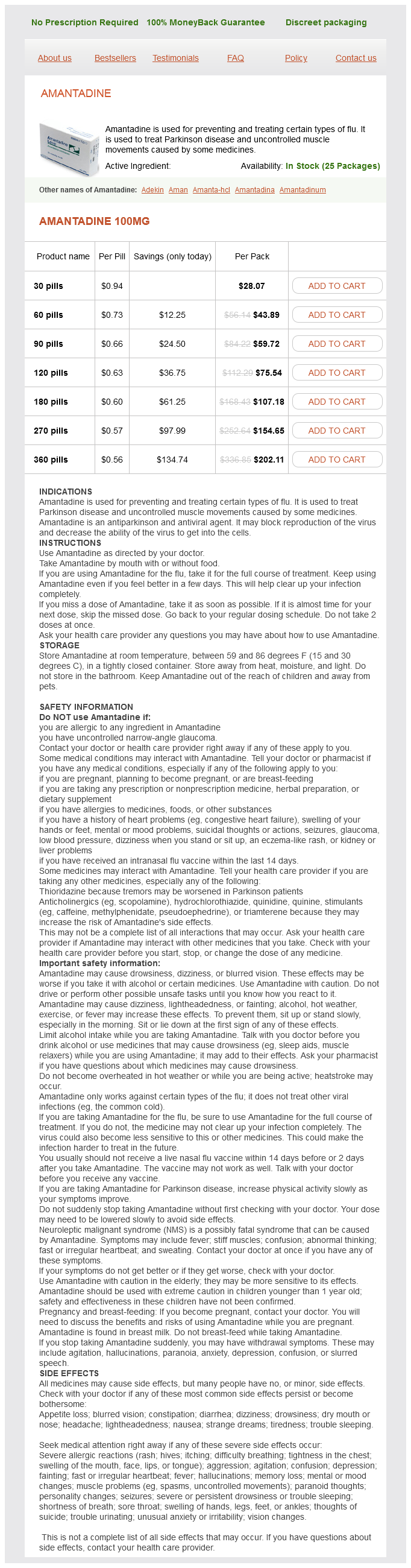
Amantadine dosages: 100 mg
Amantadine packs: 30 pills, 60 pills, 90 pills, 120 pills, 180 pills, 270 pills, 360 pills
In stock: 646
Cardiac muscle shares structural and functional characteristics with skeletal muscle and smooth muscle hiv infection chart trusted amantadine 100 mg. Both cardiac and smooth muscle cells retain their individuality, although both are in functional communication with their neighbors through gap junctions. There are three major types of muscle tissue: skeletal, cardiac, and smooth muscle. Endomysium surrounds individual fibers; perimysium surrounds a group of fibers to form a fascicle; and epimysium is dense connective tissue that surrounds the entire muscle. Three types of skeletal muscle fibers are distinguished based on contractile speed, enzymatic velocity, and metabolic profile. It is composed of precisely aligned myofilaments: myosin-containing thick filaments and actin-containing thin filaments. The arrangement of thick and thin filaments gives rise to the density differences that produce the cross-striations of the myofibril. The light-staining isotropic I band contains mainly thin filaments attached to both sides of the Z line, and the dark-staining anisotropic A band contains mainly thick filaments. Z lines between sarcomeres contain an actin-binding protein (-actinin) and Z matrix proteins. The actomyosin cross-bridge cycle represents a series of coupled biochemical and mechanical events between myosin heads and actin molecules that lead to muscle contraction. There are five recognizable stages of the cycle: attachment, release, bending, force generation, and reattachment. Regulation of muscle contraction involves Ca2, sarcoplasmic reticulum, and the transverse tubular system. The sarcoplasmic reticulum forms enlarged terminal cisternae that serve as reservoirs for Ca2. Their plasma membrane contains an abundance of gated Ca2 -release channels (ryanodine receptors [RyR1]). The transverse tubules (T tubules) are formed by invaginations of the sarcoplasm that penetrate the muscle fiber between adjacent terminal cisternae. Triads are located at the junction between A and I bands (two per each sarcomere). The depolarization of the T tubule membrane triggers the release of Ca2 from the terminal cisternae to initiate muscle contraction by binding to troponintropomyosin complex. The neuromuscular junction (motor end plate) is the contact area of the axon endings with muscle fiber.
American corn mint (Japanese Mint). Amantadine.
Source: http://www.rxlist.com/script/main/art.asp?articlekey=96610
Schematic drawing of bone marrow shows several features: erythroblastic islets engaged in the formation of erythrocytes hiv infection rates in pakistan amantadine 100 mg online, megakaryocytes discharging platelets into the sinusoids, endothelial cells (pink) resting on a basal lamina (dark red) that is absent where blood cells are entering the sinusoids, adventitial or reticular cells (blue) extending from the basal lamina into the hemopoietic compartment, and dispersed adipose cells. Photomicrograph of bone marrow section stained with H&E shows active hemopoietic centers in a close proximity to bone marrow sinusoids. Bone marrow not active in blood cell formation contains predominately adipose cells, giving it the appearance of adipose tissue. It is the chief form of bone marrow in the medullary cavity of adult bones that are no longer hemopoietically active, such as the long bones of the arms, legs, fingers, and toes. Even in hemopoietically active bone marrow in adult humans-such as that in the ribs, vertebrae, pelvis, and shoulder girdle-about half of the bone marrow space is occupied by adipose tissue and half by hemopoietic tissue. The yellow bone marrow retains its hemopoietic potential, however, and when necessary, as after severe loss of blood, it can revert to red bone marrow, both by extension of the hemopoietic tissue into the yellow bone marrow and by repopulation of the yellow bone marrow by circulating stem cells. Bone marrow examination is essential for diagnosis and management of many disorders of the blood and bone marrow. Examination of bone marrow aspirate and bone marrow core needle (trephine) biopsy is essential for the diagnosis of bone marrow disorders. Both methods are complementary and provide a comprehensive evaluation of the bone marrow. There are several indications for bone marrow examination: unexplained anemia (low erythrocyte counts), abnormal peripheral blood smear morphology, diagnosis and staging of hematological malignant disorders. Usually, the final diagnosis is based on a combination of clinical findings and several diagnostic procedures, including peripheral blood examination, bone marrow aspirate and core biopsy, and other specific tests. In bone marrow aspiration, a needle is inserted through the skin until it penetrates bone. The preferred anatomical site for a bone marrow biopsy is the posterior part of the iliac crest (hip bone). A small amount of bone marrow is obtained by applying negative pressure with a syringe attached to the needle. The aspirate is then spread as a smear on a glass slide and the specimen is examined with the microscope to examine individual cell morphology. In bone marrow core biopsy, intact bone marrow is obtained for laboratory analysis. Usually a small incision is made in the skin to allow the biopsy needle to pass into the bone. The biopsy needle is advanced through the bone with a rotating motion (similar to a corkscrew movement through a cork) and later pulled out with a small, solid piece of bone marrow inside. After the needle is withdrawn, the core sample is removed from the needle and processed for routine H&E slide preparation. The core biopsy specimen obtained in this procedure provides for analysis of bone marrow architecture.
The leukocytes and platelets are contained in a narrow over the counter antiviral cream proven 100 mg amantadine, light-colored layer between the erythrocytes and plasma called the buffy coat. Albumin is the main protein constituent of the plasma, accounting for approximately half of the total plasma proteins. Albumin is responsible for exerting the concentration gradient between blood and extracellular tissue fluid. This major osmotic pressure on the blood vessel wall, called the colloid osmotic pressure, maintains the correct proportion of blood to tissue fluid volume. If a significant amount of albumin leaks out of the blood vessels into the loose connective tissue or is lost from the blood to urine in the kidneys, then the colloid osmotic pressure of the blood decreases, and fluid accumulates in the tissues. Globulins include the immunoglobulins (-globulins), the largest component of the globulin fraction, and nonimmune globulins (-globulin and -globulin). The immunoglobulins are antibodies, a class of functional immunesystem molecules secreted by plasma cells. They help maintain the osmotic pressure within the vascular system and also serve as carrier proteins for various substances such as copper (by ceruloplasmin), iron (by transferrin), and the protein hemoglobin (by haptoglobin). Nonimmune globulins also include fibronectin, lipoproteins, coagulation factors, and other molecules that may exchange between the blood and the extravascular connective tissue. In a series of cascade reactions with other coagulation factors, soluble fibrinogen is transformed into the insoluble protein fibrin (323 kDa). During conversion of fibrinogen to fibrin, fibrinogen chains are broken to produce fibrin monomers that rapidly polymerize to form long fibers. These fibers become cross-linked to form an impermeable net at the site of damaged blood vessels, thereby preventing further blood loss. With the exception of these large plasma proteins and regulatory substances, which are small proteins or polypeptides, most plasma constituents are small enough to pass through the blood vessel wall into the extracellular spaces of the adjacent connective tissue. This method differs from the usual preparation seen in the histology laboratory in that the specimen is not embedded in paraffin and sectioned. Rather, a drop of blood is placed directly on a slide and spread thinly over the surface of the slide. Plasma proteins do not possess a structural form above the molecular level; thus, when they are retained in blood vessels in the tissue block, they appear as a homogeneous substance that stains evenly with eosin in hematoxylin and eosin (H&E)stained sections. A blood clot consists mostly of erythrocytes entangled in a network of fine fibers composed of fibrin. To prevent clotting of a blood sample, an anticoagulant such as citrate or heparin is added to the blood specimen as it is obtained. Citrate binds calcium ions, which are essential for triggering the cascade of coagulation reactions; heparin deactivates the clotting factors in the plasma. For many biochemical laboratory tests, plasma and blood serum can be used interchangeably. Serum is preferred for several specific tests because the anticoagulants in plasma can interfere with the results. However, tests of clotting ability require that all coagulation factors be preserved; therefore, serum is inappropriate for these tests.
Syndromes
Additional information:
Usage: a.c.
Tags: purchase 100 mg amantadine with mastercard, purchase 100 mg amantadine overnight delivery, generic amantadine 100 mg line, 100 mg amantadine order amex
Jaffar, 48 years: Due to relatively normal gonadotropin levels, these patients will secrete estrogen and there ore can also be said to have chronic anovulation with estrogen present.
Olivier, 25 years: The 7S domain of the tetramer (called the 7S box) determines the geometry of the tetramer.
Spike, 35 years: Visible organelles include a mitochondrion, microtubules, a single profile of the surfaceconnected open canalicular system, profiles of the dense tubular system, the moderately dense granules, a single very dense granule, and glycogen particles.
Tukash, 50 years: In sections, the cells in hemopoietic compartment appear to lie in "cords" between sinusoids or between sinusoids and bone.
Tyler, 41 years: However, randomized prospective trials have shown that adjuvant chemotherapy also improves survival rates or highrisk node-negative patients (Fisher, 2004a).