-
(786) 502-2173
-
We've gone mobile!
-
Hours: By Appointment Only
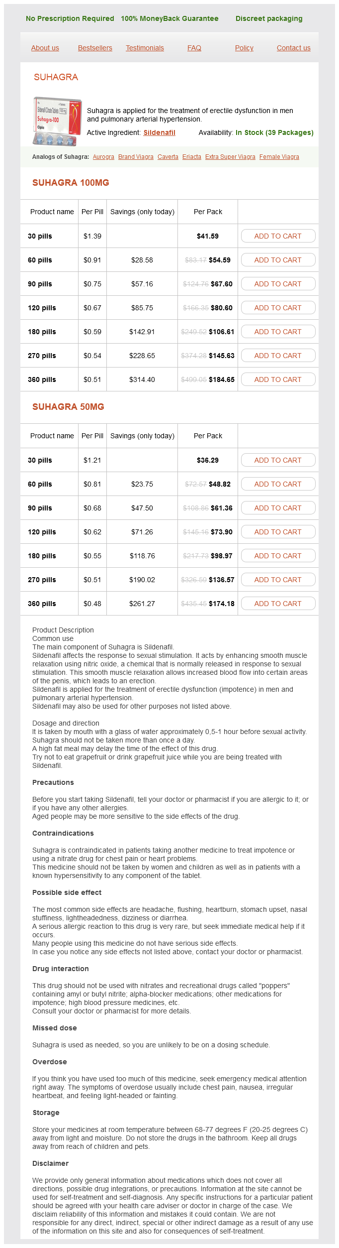
Only $0.51 per item
Suhagra dosages: 100 mg, 50 mg
Suhagra packs: 30 pills, 60 pills, 90 pills, 120 pills, 180 pills, 270 pills, 360 pills
In stock: 680
Studies have shown very good intraobserver and interobserver agreement on recognition of sphincter defects using endoanal ultrasound erectile dysfunction medication new zealand suhagra 100 mg order overnight delivery. Day three radiograph showing orally ingested radio-opaque markers mainly concentrated in the descending colon; few markers seen in the rectum. Findings of residual markers in proximal portions of the ascending, transverse, and descending colon are consistent with slow transit constipation. The findings of markers trapped in the rectum after normally transiting through the proximal colon are consistent with outlet obstruction constipation. Frequently, patients with chronic constipation will have normal colonic transit marker studies and other pathophysiologic explanation should be sought. Anal sphincter complex structures can be visualized with multiple imaging modalities. Endoanal ultrasound has become the gold standard to evaluate the anal sphincter complex and can assist in the evaluation of fecal incontinence. The most common clinical use of endoanal ultrasound is in assessment of the anal sphincter integrity in patients with fecal incontinence. The simplest method is that 20 markers are swallowed on day 0 with the follow-up x-ray taken on day five. In patients with normal colonic transit, 80% of markers should be expelled by day five. Endoanal ultrasound demonstrating anterior external and internal anal sphincter defects. Recent data suggest that injury in the puborectalis or pubovisceral muscles can result from vaginal delivery and lead to pelvic floor dysfunction. Transperineal and translabial pelvic floor ultrasound is an active area of research with promising future clinical utility in imaging the pelvic floor musculature. Defects in the levator ani muscles have been found in women after vaginal delivery and reported at higher prevalence in women with fecal incontinence and prolapse. The images are obtained in axial plane with T2-weighted fast spin and T1-weighted spin sequences. Muscle defects can be seen as disruption of the anal sphincter ring or hyposignal scarring, whereas atrophy appears as thinning or fatty replacement. Dynamic Imaging Fluoroscopic Evacuation Proctography (Defecography) Fluoroscopic evacuation proctography, also known as defecography, is a morphologic and functional examination of the anorectum and the pelvic floor, which allows for realtime physiologic assessment of defecation. Normal pelvic floor hiatus on transperineal ultrasound with anatomic structures identified. This image is from a vaginally parous woman with no visible injury to the puborectalis muscle or changes in the pelvic floor hiatus.
Citrus Peel extract (Grapefruit). Suhagra.
Source: http://www.rxlist.com/script/main/art.asp?articlekey=96909
Pressure on the posterior wall of the vagina erectile dysfunction doctors naples fl 50 mg suhagra buy, directed toward the rectum, may facilitate identification of rectal prolapse. Assessment of the anal sphincter may also be performed including evaluation of anal tone, squeeze, and symmetry. The clinical examination enables documenting the presence or absence of prolapse, but is not accurate in identifying the location of the connective tissue defect or presence of an enterocele or sigmoidocele. A woman with defecatory dysfunction and pelvic organ prolapse may benefit from further testing. Defecography provides a two-dimensional view of the efficiency of anorectal emptying during simulated defecation and quantification of rectal parameters. Prior to the test, a woman ingests diluted barium, the rectum is filled with a barium paste that has the consistency of soft stool and the barium gel is placed in the vagina. Diagnostic categories obtained are internal procidentia, rectocele, enterocele, sigmoidocele, descending perineum, and the functional description of spastic pelvic floor. Some degree of rectocele is present in most symptomatic women and up to 20% of asymptomatic women. Descent of the perineal body and descent of the bowel into the rectovaginal space can be visualized and graded (Table 13-3). Retention of more than 10% of the barium following defecation is referred to as barium trapping. Paradoxical contraction of the puborectalis and rectal intussusception may be diagnosed with this functional test. The disadvantages include exposure to radiation and inability to image soft tissues. The technique in the sitting position simulates the functional testing achieved with defecography with the added advantages of superb soft tissue imaging, large field of view, avoidance of ionizing radiation, direct multiplanar capability, and high temporal resolution. At this time, a standardized method of establishing a radiologic diagnosis of a rectocele is lacking. These questionnaires may be performed pre- and postoperatively to provide a standardized method of evaluating functional surgical outcomes. A woman who describes life-long infrequent bowel movements defined as less than one bowel movement per week and an absence of a daily urge to defecate is unlikely to be cured of her constipation with a rectocele repair. A colon transit study may be helpful in identifying patients with slow transit colon. Dietary modifications including fiber and laxatives should be encouraged in any woman whose main complaint is constipation.
Degenerative tendinitis of the rotator cuff is common erectile dysfunction pills otc purchase suhagra 100 mg without a prescription, especially in older people. These syndromes are discussed in detail later in this chapter, in relationship to the glenohumeral (shoulder) joint. Bursae around the glenohumeral (shoulder) joint, between the tendons of the rotator cuff muscles and the fibrous layer of the joint capsule, reduce friction on the tendons passing over the bones or other areas of resistance. The shape and size of the axilla vary depending on the position of the arm; it almost disappears when the shoulder joint is fully abducted. The axilla provides a passageway for vessels and nerves going to and from the upper limb. The axilla has an apex, base, and four walls, three of which are muscular: · the apex of the axilla is the cervico-axillary canal, the passageway between the neck and the axilla. The clavipectoral triangle is bounded by the clavicle superiorly, the deltoid laterally, and the clavicular head of pectoralis major medially. When the arm is abducted and then adducted against resistance, the two heads of the pectoralis major are visible and palpable. As this muscle extends from the thoracic wall to the arm, it forms the anterior axillary fold. Digitations of the serratus anterior appear inferolateral to the pectoralis major. The coracoid process of the scapula is covered by the anterior part of deltoid; however, the tip of the process can be felt on deep palpation in the clavipectoral triangle. The area formed by the superior border of latissimus dorsi, the medial border of the scapula, and the inferolateral border of the trapezius is called the triangle of auscultation. This gap in the thick back musculature is a good place to auscultate the posterior segments of the lungs with a stethoscope. When the scapulae are drawn anteriorly by folding the arms across the thorax and the trunk is flexed, the triangle of auscultation enlarges. The teres major forms a raised oval area on the inferolateral third of the posterior aspect of the scapula when the arm is adducted against resistance. The posterior axillary fold is formed by the teres major and the tendon of the latissimus dorsi. The base of the axilla is formed by the concave skin, subcutaneous tissue, and axillary (deep) fascia extending from the arm to the thoracic wall forming the axillary fossa (armpit). The anterior wall of the axilla is formed by the pectoralis major and minor and the pectoral and clavipectoral fascia associated with them. The posterior wall of the axilla is formed chiefly by the scapula and subscapularis on its anterior surface and inferiorly by the teres major and latissimus dorsi.
Syndromes
Additional information:
Usage: b.i.d.
Tags: suhagra 100 mg on-line, 100 mg suhagra with visa, suhagra 100 mg order, effective 100 mg suhagra
Ressel, 27 years: So far there are few studies in the area of pelvic pain including effective diagnostic, prevention, and therapeutic interventions. Physical Examination Findings on digital rectal examination may include anal stricture or mass, paradoxical contraction of the puborectalis, nondescent of perineum and rectocele/ enterocele.
Hjalte, 49 years: Pelvic tumors Glycosuria Genetic Hypoestrogenic state Sexual intercourse Spermicides Genetic factors have been postulated to increase the risk of recurrent infection. Synovial sarcoma in older patients: clinicopathological analysis of 32 cases with emphasis on unusual histological features.
Mazin, 44 years: Articular cartilage Head of femur Epiphysial plate Femur Int tervertebral disc Body of vertebra Anterior view Primary cartilaginous (Synchondrosis) Lateral view Secondary cartilaginous (Symphysis) In cartilaginous joints, articulating bones are united by fibrocartilage or hyaline cartilage. Diagnosis of anal sphincter tears to prevent fecal incontinence: a randomized controlled trial.
Kalesch, 36 years: Osteoporosis is characterized by a net demineralization of bones and results from a disruption of the normal balance of calcium deposition and resorption. Abdominal and/or vaginal repairs for correction of prolapse are often performed concomitantly with a Burch colposuspension.
Rufus, 34 years: In the inferior part of the femoral triangle, the femoral vein receives the profunda femoris vein, the great saphenous vein, and other tributaries. The thoracic aorta lies posterior to the root of the left lung, the pericardium, and the esophagus.