-
(786) 502-2173
-
We've gone mobile!
-
Hours: By Appointment Only
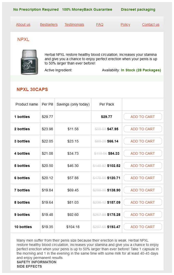
Only $20.56 per item
NPXL dosages: 30 caps
NPXL packs: 1 bottles, 2 bottles, 3 bottles, 4 bottles, 5 bottles, 6 bottles, 7 bottles, 8 bottles, 9 bottles, 10 bottles
In stock: 746
Involvement of the spine most commonly manifests as vertebral osteomyelitis including punched-out and permeative lesions within the vertebral bodies herbs you can smoke 30 caps npxl order mastercard. The treatment of spinal coccidioidomycosis combines medical management with antifungal medications and surgery including spinal fusion. There is swelling and high signal intensity of the anterior aspect of the intervertebral disk with mild prevertebral soft tissue edema. A: Photograph of a sagittal section of the thoracic spine shows complete destruction of two of the vertebral bodies including the intervertebral disk. The tuberculous abscess has extended anteriorly and posteriorly, causing compression of the spinal cord (arrow). B: Photomicrograph of a tuberculous granuloma shows central caseation necrosis surrounded by inflammatory tissue (H&E, original magnification ×80). C: A highpower view shows a characteristic for tuberculosis Langhans giant cell with a horseshoe-shaped distribution of nuclei (arrow), surrounded by a palisade of epithelioid histiocytes and chronic inflammatory cells including lymphocytes and plasma cells (H&E, original magnification ×450). A: Anteroposterior radiograph of the thoracic spine in a 50-year-old man shows narrowing of the T8T9 disk space, associated with a paraspinal mass on the left side (open arrows). B: Lateral conventional tomogram shows destruction of the disk and extensive erosions of the inferior aspect of the body of T8 and the superior end plate of T9. Lateral radiograph of the lower thoracic spine of a 12-year-old boy with long-standing pulmonary and spinal tuberculosis shows severe kyphosis with a gibbous formation at the site of a collapsed vertebra, a common complication of this condition. Anteroposterior radiograph of the pelvis in a 35-year-old woman with spinal tuberculosis shows an oval radiodense mass with spotted calcifications overlapping the medial part of the ilium and right sacroiliac joint (right psoas muscle) (arrows). A 39-year-old man with a history of pulmonary tuberculosis had neurologic symptoms of spinal cord compression. A: Anteroposterior radiograph of the lower thoracic spine shows minimal disk space narrowing at T9T10 and a large left paraspinal mass (arrowheads). B: A myelogram shows complete obstruction of the flow of contrast in the subarachnoid space at the level of the disk infection (arrows). Positron emission tomography as a diagnostic tool in infection: present role and future possibilities. The use of monoclonal antibodies and antibody fragments in the imaging of infectious lesions. Embolic osteomyelitis of the spine as a complication of infection of the urinary tract. Infectious granulomas of bones and joints, with special reference to coccidioidal granuloma. Tuberculous spondylitis and pyogenic spondylitis: comparative magnetic resonance imaging features. Coccidioidomycosis: a descriptive survey of a reemerging disease clinical characteristics and current controversies. Septic arthritis with negative bacteriological findings in adult native joints: a retrospective study of 74 cases. Radiographic findings in early acquired syphilis: case report and critical review.
Ginseng root (Ginseng, Panax). NPXL.
Source: http://www.rxlist.com/script/main/art.asp?articlekey=96961
Infants and small children involved in motor-vehicle crashes can be transported in their car seats as long as they have no apparent injury and the device is not damaged herbs de provence walmart buy npxl 30 caps without prescription. Recently released guidelines from the National Athletic Trainers Association recommend helmet and pad removal prior to transport, when there is a suspected unstable spine injury. The four main reasons to consider field athletic helmet removal are · Face mask cannot be removed in a timely fashion · Airway cannot be controlled due to the design of the helmet and chin strap · Helmet and chin straps do not hold the head securely · Helmet prevents stabilization for transport in an appropriate position When removing an athletic helmet, it is imperative to cut the chin strap and not attempt to unsnap or unbuckle the device. Notice that the helmet flexes the neck in a patient who is not wearing shoulder pads. Not only is this done routinely with helmet removal, but it is also done when faced with the inability to maintain neutral cervical-spine alignment (often due to ill-fitting shoulder pads), when you are unable to secure the athlete to the stretcher or spine board, and when you need access to the chest for resuscitation efforts. Most shoulder pads can be removed by cutting the axillary straps and the laces on the front of the appliance, opening the appliance from the core outward (like a clam shell), and sliding the appliance out from under the athlete. Motorcycle helmets often are designed with a continuous solid face guard that limits airway access. Those helmets are not custom designed and frequently are poorly fitted to the patient. The motorcycle helmet can make it difficult to stabilize the neck in a neutral position, obstruct access to the airway, and hide injuries to the head or neck. It should be removed in the prehospital setting, using the techniques described in Chapter 12. Very Large or Obese Patients Very large or obese patients might not fit appropriately in standard equipment. In the absence of such resources, emergency care providers may improvise, such as using sheets of plywood with head cushions or towel rolls to stabilize the spine. This should be done with caution because the weight capacity and safety of such improvised equipment may not be known. Any immobilized large patient should have the head elevated to prevent respiratory compromise. Larger patients who do not have spine injuries should be transported in an upright position, which is typically the position of comfort. In cold climates, patients in bulky warm clothing will need to be snugly secured to prevent excessive movement. Cervical collars should not be used because they will prevent continued examination of the wound site and could compromise the airway in wounds with expanding hematomas or subcutaneous air. Therefore, for patients with such injuries, it may be wise to avoid collars and use instead manual stabilization and head cushion devices or blanket rolls for cervical motion restriction, if clinically indicated. Case Presentation (continued) You are at the scene of a two-vehicle collision involving a car and a small truck that has rolled over. You are asked to check on the driver of the overturned truck, who extricated himself and is standing, talking to a police officer. Because he is speaking, answering questions, and walking, you conclude that his primary survey is intact.
Rarely harbs cake nyc discount npxl 30 caps fast delivery, extensive calcifications or ossifications resembling an osteoid matrix or bone can be present. This presentation may lead to erroneous diagnosis including soft tissue osteosarcoma or chondrosarcoma, synovial chondromatosis, myositis ossificans, or tumoral calcinosis. A: Monophasic variant exhibits dense arrangements of spindle cells resembling fibrosarcoma (H&E, original magnification ×50). D: Calcifying type of the tumor exhibits extensive mineralization (H&E, original magnification ×100). E: At high magnification, observe uniformity of the spindle cells with only mild pleomorphism surrounding areas of mineralization (H&E, original magnification ×400). Lateral radiograph of the left ankle of a 71-year-old woman shows a large calcified mass located in the soft tissues anteriorly to the Achilles tendon, not affecting the adjacent bones. Scintigraphic evaluation reveals increased uptake of radiopharmaceutical agent on blood flow and blood pool images consistent with increased vascularity of these tumors. The lesion was usually heterogeneous on T2weighted images and was clearly delineated from surrounding tissues. Fortyfour percent of the cases had a high signal on both T1- and T2-weighted sequences, consistent with hemorrhage within the tumor. Several investigators consider so-called "triple signal intensity" sign, because of a combination of cystic and solid elements, fibrous tissue, hemorrhage, and hemosiderin deposition, as the most characteristic for this tumor. Some multilobulated tumors may contain septa and fluidfluid levels, creating the "bowl of grapes" sign. Treatment includes a wide local resection, followed with adjuvant chemotherapy with combination of Adriamycin, cisplatin, vincristine, doxorubicin, and ifosfamide. Postoperative radiation therapy is reserved for the patients in whom surgical intervention was not able to ascertain clear margins of resection. It may arise as a primary synovial tumor, or it may develop as a malignant transformation of synovial (osteo)chondromatosis. The concept of malignant degeneration of synovial chondromatosis is still controversial and the entity is rare, with fewer than 40 well-documented cases on record. These malignancies show a slight predominance in men, and patients range in age from 25 to 70 years. The symptoms include pain and swelling, with duration in most patients exceeding 12 months. In patients with primary synovial (osteo)chondromatosis, malignant transformation to synovial chondrosarcoma should be clinically suspected if there is development of soft tissue mass at the site of the affected joint. Radiologically, the presence of chondroid calcifications within the joint, destruction of the adjacent bones, and a soft tissue mass are highly suggestive of a synovial chondrosarcoma.
Syndromes
Additional information:
Usage: q.h.
Tags: cheap npxl 30 caps buy, cheap 30 caps npxl free shipping, buy npxl 30 caps on-line, discount npxl 30 caps buy online
Irmak, 49 years: They are located chiefly in the mid-zones, paravertebral gutters and over the central tendons of the diaphragm.
Eusebio, 22 years: There is slightly increased signal in the critical zone of the supraspinatus tendon.
Cruz, 58 years: Investigations Duplex scanning: B-mode scan and Doppler ultrasonic velocitometry: method of choice for assessing degree of carotid stenosis.
Tempeck, 48 years: Herpesvirus 8 inclusions in primary effusion lymphoma: report of a unique case with T-cell phenotype.
Shakyor, 52 years: Foci of mesothelial hyperplasia, associated with a borderline serous tumor, may be wrongly interpreted as indicating the serous tumor has an invasive component.