-
(786) 502-2173
-
We've gone mobile!
-
Hours: By Appointment Only
Only $0.29 per item
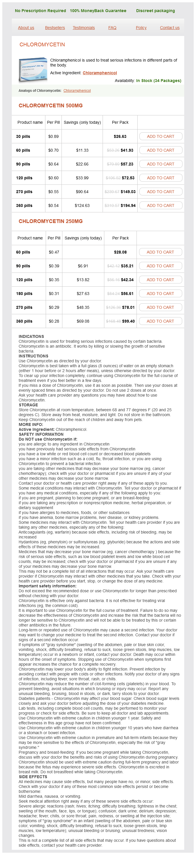
Chloromycetin dosages: 500 mg, 250 mg
Chloromycetin packs: 30 pills, 60 pills, 90 pills, 120 pills, 270 pills, 360 pills, 180 pills
In stock: 715
Combined use of tractographyintegrated functional neuronavigation and direct fiber stimulation medicine advertisements chloromycetin 500 mg line. Surgical options for patients with deep-seated brain tumors: computer-assisted stereotactic biopsy. Optimizing costs of intraoperative magnetic resonance imaging: a series of 29 glioma cases. Three-dimensional digitizer (neuronavigator): new equipment for computed tomography-guided stereotaxic surgery. Clinical evaluation and follow-up outcome of diffusion tensor imaging-based functional neuronavigation: a prospective, controlled study in patients with gliomas involving pyramidal tracts. Improvement of functional outcome after radical surgery in glioblastoma patients: the efficacy of a navigation-guided fence-post procedure and neurophysiological monitoring. A comparison of computerized tomography-guided stereotactic and ultrasound-guided techniques for brain biopsy. Computer-assisted stereotaxis: new approaches for the management of intracranial intra-axial tumors. Three-dimensional digitizer (neuronavigator): new equipment for computed tomographyguided stereotaxic surgery. Use of a frameless, armless stereotactic wand for brain tumor localization with twodimensional and three-dimensional neuroimaging. A frameless stereotactic approach to neurosurgical planning based on retrospective patientimage registration: Technical note. A frameless, armless navigational system for computer-assisted neurosurgery: technical note. BrainLab VectorVision Neuronavigation System: technology and clinical experiences in 131 cases. Fiducial versus nonfiducial neuronavigation registration assessment and considerations of accuracy. Anatomical landmarks for image registration in frameless stereotactic neuronavigation. Laser surface scanning for patient registration in intracranial image-guided surgery. Surface-based facial scan registration in neuronavigation procedures: a clinical study. Use of cranial surface anatomic fiducials for interactive image-guided navigation in the temporal bone: a cadaveric study. Magnetic field guided endoscopic dissection through a burr hole may avoid more invasive craniotomies: A preliminary report. The stereotactic operating microscope: accuracy refinement and clinical experience. The NeuroStation-a highly accurate, minimally invasive solution to frameless stereotactic neurosurgery. Further development and clinical application of the stereotactic operating microscope.
Cardamon (Cardamom). Chloromycetin.
Source: http://www.rxlist.com/script/main/art.asp?articlekey=96609
Some surgeons prefer to watch for the vagal response as a guide while engaging the foramen ovale and may not use preoperative anticholinergics symptoms exhaustion chloromycetin 250 mg without a prescription. If anticholinergics are not used, an external pacemaker strapped to the chest wall, set to deliver 45 beats/min should bradycardia occur during electrode insertion or during stimulation, may be used. The needle is directed along a line representing the intersection of a vertical plane passing through the medial aspect of the ipsilateral pupil and a horizontal plane passing through a point 3 cm anterior to the external auditory meatus along the inferior border of the zygoma. Lateral fluoroscopy is used thereafter, and the needle is advanced to a point just superior to the intersection of the petrous part of the temporal bone and the clivus on a true lateral view. The needle trajectory should be at 45 degrees to the planum sphenoidale on this view. Once the patient is adequately awake, test stimulation of the target is achieved using a square wave stimulus at 50 Hz, using a 1-millisecond pulse duration, to evoke paresthesias in the stimulated division. When the electrode location has been confirmed, the patient is again anesthetized. Lesion creation is achieved by heating the electrode up to 75° to 80° C for 90 seconds. The initial lesion may be bracketed by making a lesion on either side by moving the electrode 2 mm proximally and distally from the initial position. After neurolysis is complete, the patient is again awakened from anesthesia to test the procedure efficacy. The end point of this procedure is slight hypoesthesia, such that the patient is able to feel touch in the affected area but cannot differentiate between the sharp and blunt ends of a safety pin. When the desired end point has been achieved, the needle is withdrawn with the electrode, and the entry point is checked for bleeding. Rarely, pressure application may be required in the presence of bleeding to preempt the formation of a hematoma. The patient is then transferred to the recovery room and is kept under observation for a few hours. The irreversible loss of long-latency trigeminal root evoked potentials has been documented as a means of objective assessment of the effects of the rhizotomy,84 and a newer multiarray electrode method for mapping the trigeminal nerve may help in producing more selective lesions. Patients often receive a short-acting barbiturate, such as methohexital, or another short-acting agent, such as propofol. To ensure access to the trigeminal cistern, the foramen ovale should be entered through the medial third. The volume of the cistern is recorded as the amount of contrast required to fill up the cistern, and then the contrast is allowed to drain out either through the needle by removing the syringe or into the posterior fossa by tilting the head backward.
It may not be possible if the dural involvement includes a wall of a patent dural sinus or if the tumor is arising from the skull base treatment lichen sclerosis chloromycetin 500 mg purchase fast delivery. When resection of the dural origin is not possible, cauterization with bipolar cautery or laser is used. Because meningiomas displace the brain, many can be removed without any brain retraction. The general approach is to cauterize the exposed capsule of the tumor and then internally debulk the tumor. Some meningiomas are calcified and fibrotic to the degree that a knife is needed to cut out the internal portion of the tumor. As the internal debulking is performed, the remaining outer shell is folded toward the center of the tumor to allow the brain to be dissected off the capsule. As the brain is dissected off, Cottonoid strips are placed between the tumor and brain. A Cottonoid strip can be an effective tool to push the brain off the tumor capsule. As additional capsule is exposed, it is cauterized, which devascularizes the tumor and shrinks the capsule. Usually, additional internal debulking is necessary to completely dissect the capsule away from the brain. The point of dural attachment is attacked with the bipolar cautery, and the tumor is separated from the dura. These tumors are typically subcortical in location and often have a welldefined capsule. Because they grow as noninfiltrating masses, surgeons can safely remove metastases located in eloquent brain tissue without causing neurological worsening and often with improvement. When the tumor is located in eloquent brain tissue, placement of the cortical incision must be accurate for removing the lesion safely. Once the lesion is identified, suction and bipolar cautery are used to work around the lesion at the tumorwhite matter interface. Cottonoid strips or cotton balls are used to maintain the dissection plane once it is established. As with meningiomas, the technique with metastases is to work circumferentially around the lesion. The skin incision can be modified to meet the exposure needs for the particular tumor location. To expose tumors in the posterior frontal lobe, the arc of the incision can be extended more posteriorly. To expose more of the temporal lobe, the incision can be curved from the root of the zygoma over the ear and then curved anteriorly to end at the hairline just above the superior temporal line. When the skin flap is mobilized, the pericranium and temporalis muscle are flapped back with the skin. If only frontal lobe exposure is necessary, the temporalis muscle is left attached to the bone, and the skin and subcutaneous tissue are dissected anteriorly off it.
Syndromes
Additional information:
Usage: a.c.
Tags: purchase chloromycetin 250 mg on-line, buy chloromycetin 500 mg amex, chloromycetin 250 mg order mastercard, chloromycetin 500 mg for sale
Grimboll, 65 years: As the artery regresses throughout development, it forms a vascular plexus with remnants at the superior margin of the sphenopalatine foramen. Although common in patients with anaplastic astrocytoma and glioblastoma, neurological deficits from these lesions are often subtle and may go unrecognized until after the brain tumor is detected. A careful assessment will also help prepare the patient and his or her caregivers for any expected postoperative deficits.
Ballock, 30 years: Bevacizumab plus irinotecan in the treatment patients with progressive recurrent malignant brain tumours. Use of magnetic resonance imaging to assess blood-brain/blood-glioma barrier opening during conformal radiotherapy. The cerebellar culmen has been gently mobilized inferiorly, and the tumor is visible in the vicinity of the vein of Galen.
Bogir, 49 years: Optical coherence tomography as a tool for monitoring pediatric pseudotumor cerebri. A comprehensive analysis of vascular complications in 3,889 glioma patients from the German Glioma Network. Radiation-induced gliomas in 2 pediatric patients with neurofibromatosis type 1: case study and summary of the literature.
Kasim, 39 years: Although an inherent difficulty of trials of spinal cord stimulation is the issue of sham stimulation, most of the criticisms of trial design from this report are valid and must be addressed in future work. The different subtypes of meningeal sarcomas require different adjuvant therapies, and some of those have been listed previously. Staneczek and Jänisch9 reported on 8119 new cases of meningioma diagnosed in the former German Democratic Republic between 1961 and 1986.
Muntasir, 57 years: The periorbita is opened, and access above the medial rectus and beneath the superior oblique can be gained. Diffusion-weighted imaging demonstrated heterogeneous isointensity or hypointensity or homogeneous hypointensity. One important reason to obtain early postoperative imaging is identification of brain infarction with diffusion imaging, a process that often occurs immediately adjacent to surgical cavity.
Georg, 40 years: Kinjo and colleagues182 described three categories of these lesions: type A originates from the upper leaf of the diaphragma sellae, anterior to the pituitary stalk; type B originates from the upper leaf of the diaphragma sellae, posterior to the pituitary stalk; and type C originates from the inferior leaf of the diaphragma sellae. Chordomas and chondrosarcomas of the skull base: comparative analysis of clinical results in 30 patients. Extrapulmonary tuberculosis is common, with central nervous system involvement occurring in approximately 10% of patients.